Immune regulation of behavior
Understanding circuits between the immune and nervous system that shape pain and sensory experience.
Understanding circuits between the immune and nervous system that shape pain and sensory experience.
Immune cells and peripheral sensory neurons occupy the same tissues, and the signals immune cells release are sensed directly by the neurons that encode pain and itch. Using genetic tools, in vivo imaging of neuron activity, and light-sheet imaging of the whole nervous system, we aim to determine:
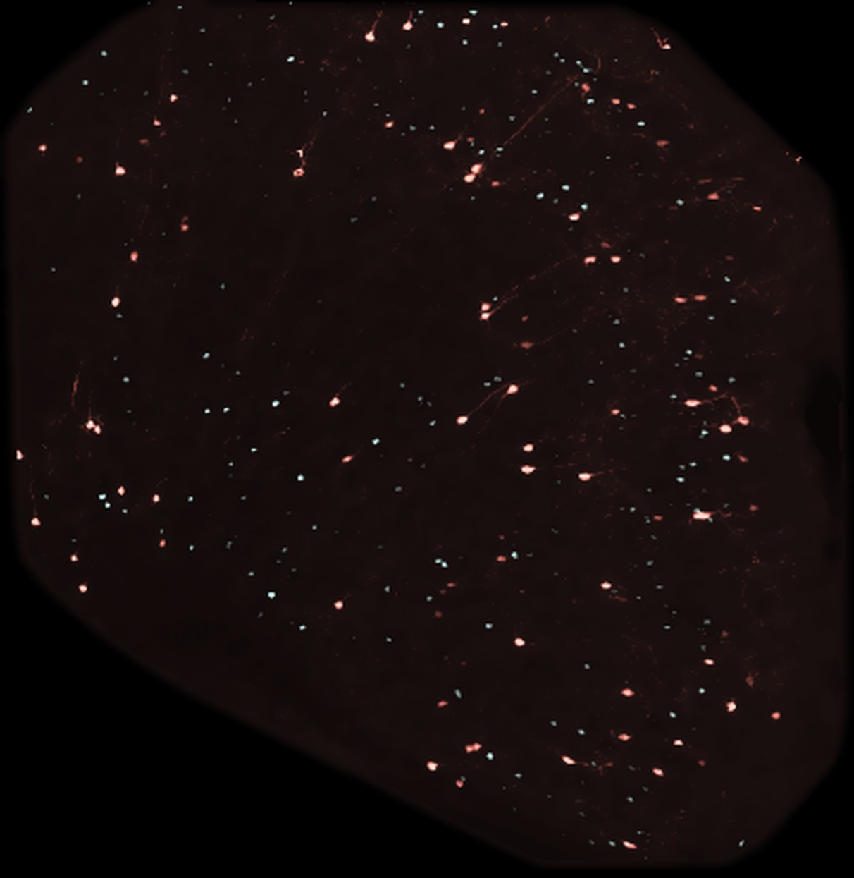 Anterior cingulate cortex
Using human patient samples, new preclinical genetic tools, and fundamental immunology, we ask how the nervous system augments or modulates inflammation. Specifically:
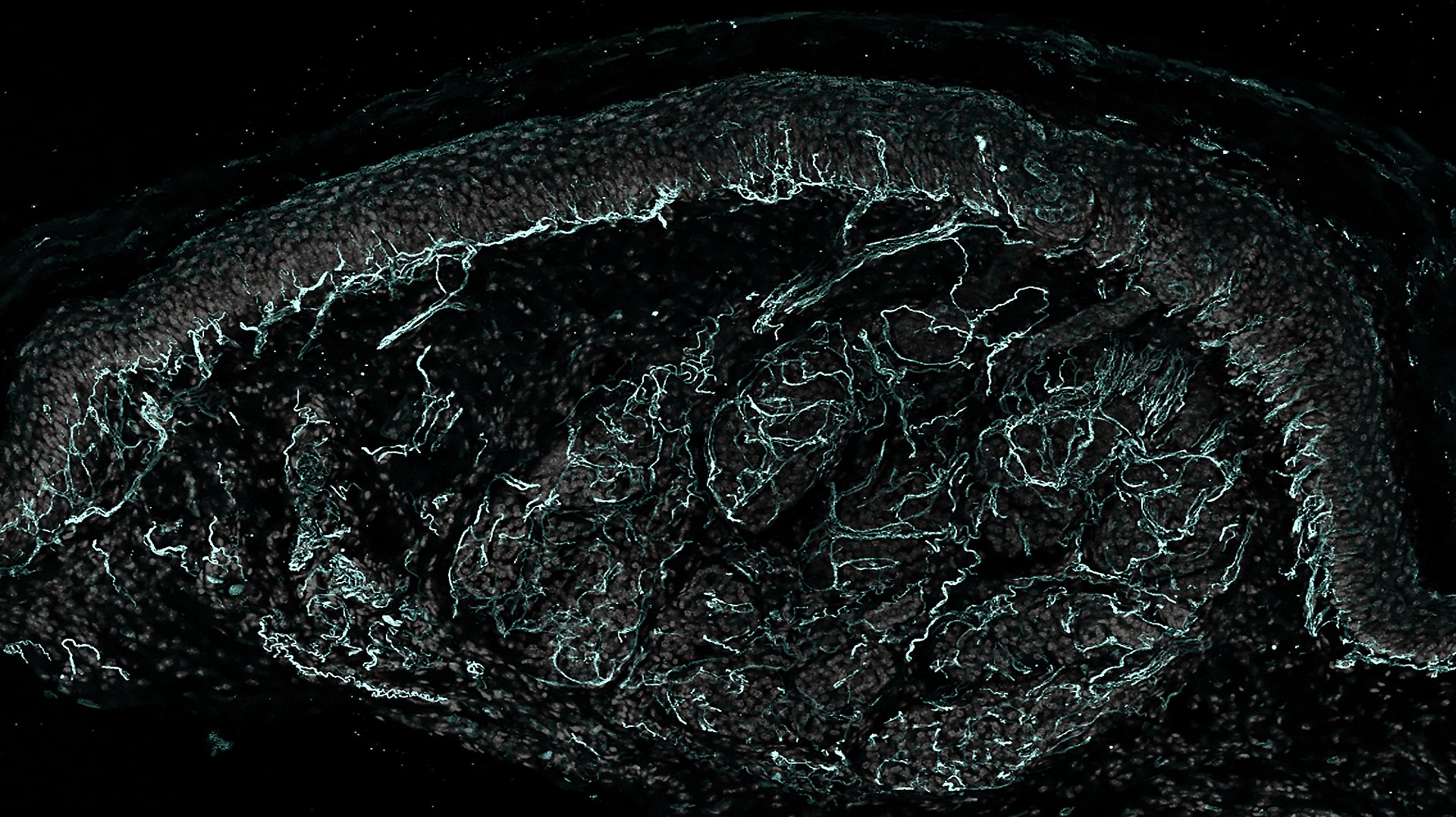 Skin
Skin
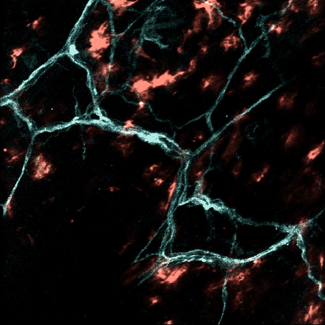 Nerve · Immune
With collaborators, we are developing new therapies for neurogenic inflammation, pain, and itch:
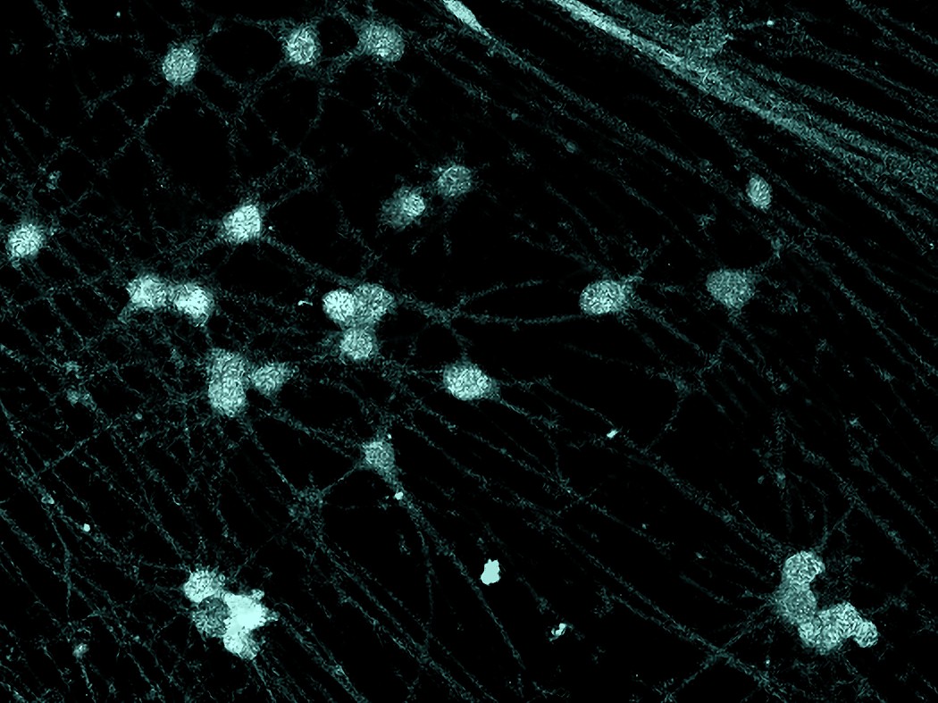 Human sensory neurons
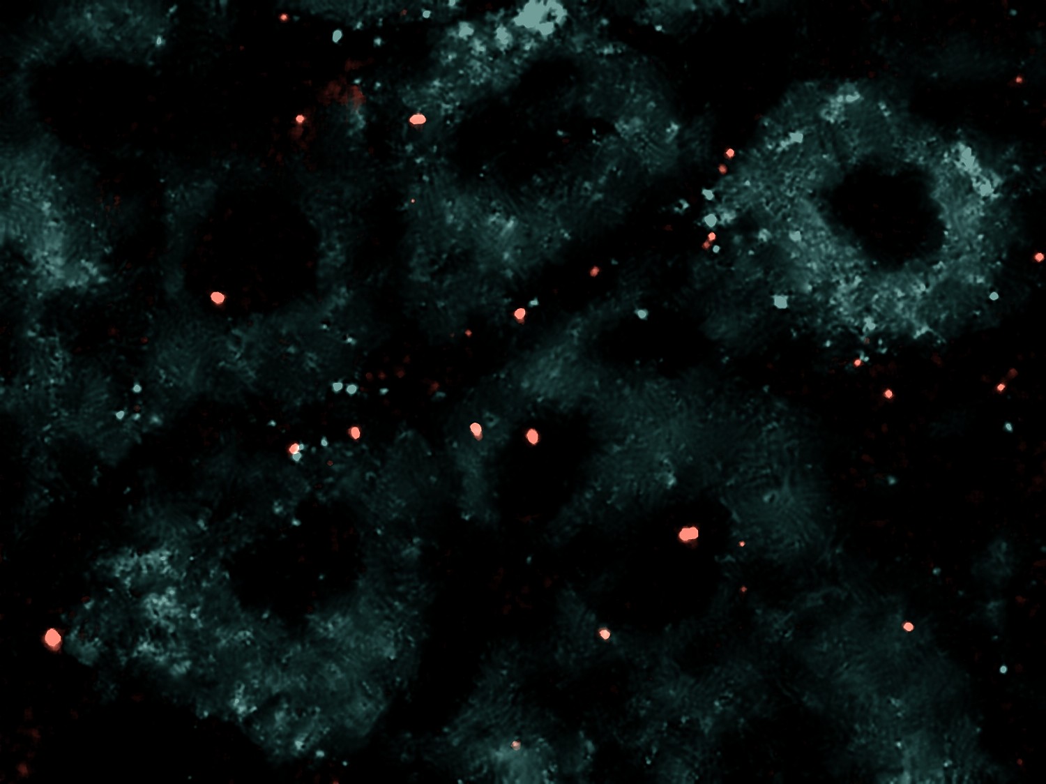 Dorsal root ganglion
Élora completed her undergraduate and graduate degrees at the Université de Sherbrooke in Quebec before joining Allan Basbaum's lab at UCSF for her postdoctoral fellowship. She studies sex differences in pain and is soon starting her own lab.
Beatriz trained at the Federal Institute of São Paulo (IFSP) and the University of São Paulo, Brazil, for her undergraduate and graduate degrees before joining the lab as a postdoctoral fellow. She studies endogenous opioid regulation of pain at tissue interfaces and is a Pew Latin American Fellow.
Daniela did her graduate work at the Brain Institute, UFRN, in Natal, Brazil, followed by postdoctoral fellowships with Sam Pleasure and Laura Cocas at UCSF. She joins the lab as a microscopy specialist, working on human sensory neuron function in response to inflammation.
Crystal did her undergraduate studies at UC Berkeley and joined the lab as a junior specialist. She works on sex-selective circuits of pain and is pursuing a career in medicine.
Sakeen trained as an MD/PhD at the University of Minnesota with Daniel Kaplan, studying immune and neural mechanisms of pathogen resistance. After internal medicine at UT Southwestern and dermatology residency at UCSF, he completed a postdoctoral fellowship in neuroanatomy with Allan Basbaum and joined the UCSF faculty, where he studied the immunobiology of pain and itch. His work is supported by the NIH, the Dermatology Foundation, the Arthritis National Research Foundation, the Burroughs Wellcome Fund, and the Sandler Foundation.
The lab is recruiting at every level. How to join →
* co-first authors
Our laboratory studies how the sensory nervous system and the immune system interact in health and disease. We are disease- and tissue-agnostic, and we aim to use the immune system as a vehicle to resolve neuroanatomy and behavior.
We are recruiting postdoctoral fellows, physician-scientists, graduate, medical, and undergraduate students, and a technician. We welcome people from all backgrounds and disciplines, with a particular interest in basic molecular and cellular biology that can be brought into the study of immunology and neuroscience. We encourage independence and work with each trainee toward their long-term goals.
Email the PI with a brief statement, one to two paragraphs, describing the questions or projects that interest you, your background, and your long-term goals, and attach a CV.
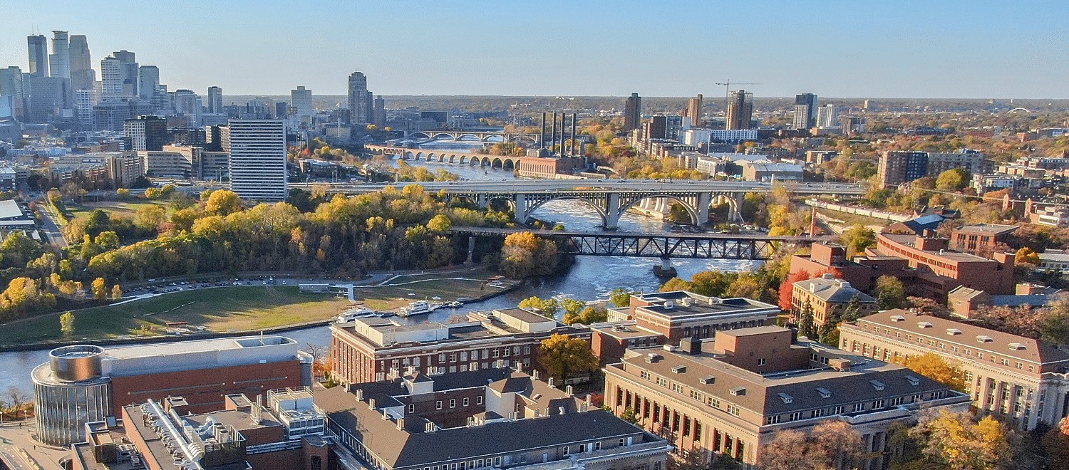
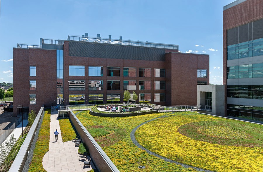
kashe007@umn.eduThe lab is currently at the Mission Bay campus of UCSF and moves to the University of Minnesota in early 2027, joining the Department of Dermatology and the Center for Immunology. We will be on the 4th floor of the Wallin Medical Biosciences Building, alongside the university's neuroscientists and the Center for Immunology.
University of California, San Francisco
Mission Bay campus
San Francisco, CA
University of Minnesota
Department of Dermatology · Center for Immunology
Wallin Medical Biosciences Building, 4th floor
2101 6th Street SE
Minneapolis, MN 55455